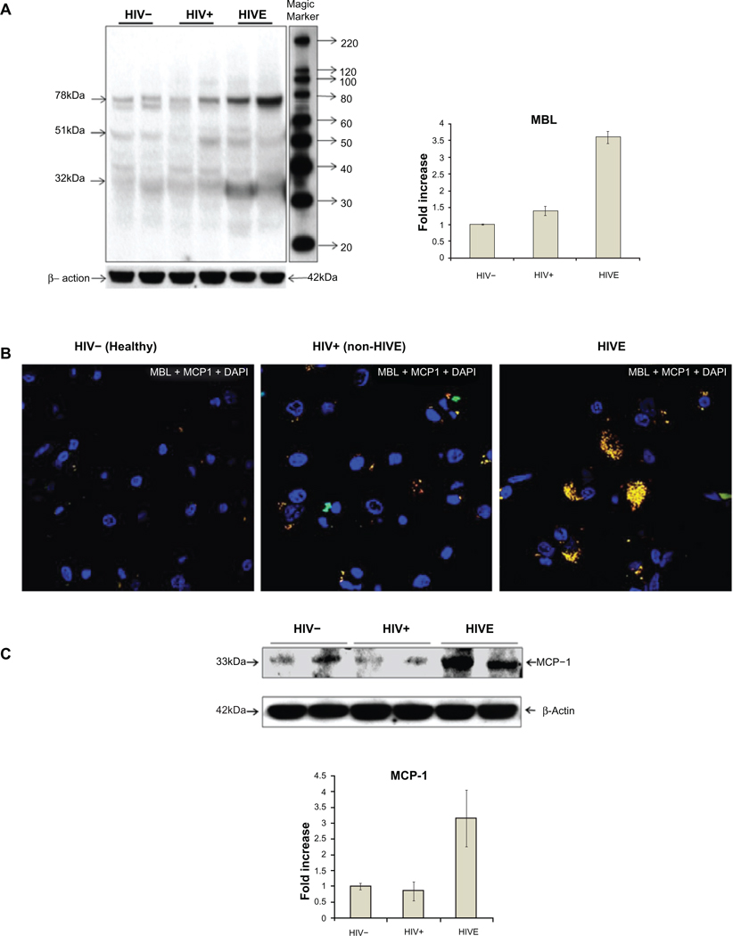
Figure 4.
Western blot analyses of mannose binding lectin (MBL) expression in post-mortem brain frontal cortex tissues from two each of HIV−, HIV+ non-HIVE, and HIVE cases (A). 32 kDa monomers, 51 kDa dimers and 78 kDa trimers of the MBL were observed. Side panel shows a 3-fold increase of MBL in HIVE vs HIV+ non-HIVE cases and bars represent the standard deviation of MBL concentration in the studied samples. Distribution of MCP-1 and its co-localization with MBL in post-mortem brain frontal cortex tissues using immunofluorescence (B) and western blot (C). Green fluorescence represents MBL, red represents MCP-1, and the yellow fluorescence represents the co-localization of MBL and MCP-1. Blue fluorescence represents the presence of an intact nucleus stained by 4’, 6-diamidino-2-phenylindole (DAPI). Method, scale, and magnification are the same as in Figure 1. Panel C shows about 3-fold increase of MCP-1 in HIVE vs non-HIVE or HIV negative cases. Bars represent the standard deviation of MCP-1 concentration in the studied samples.

