Figure 2.

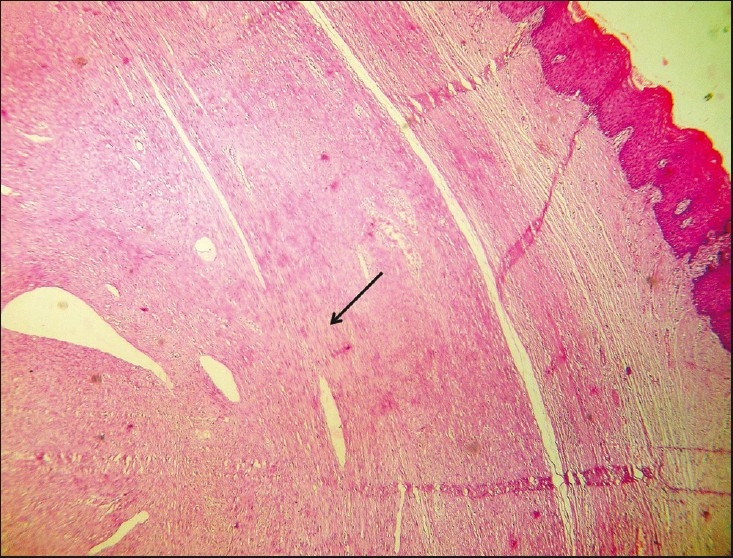
Microphotograph showing the leiomyoma (arrow) with the overlying vaginal squamous epithelium (right side) (hematoxylin and eosin stain, ×100 magnification)

Microphotograph showing the leiomyoma (arrow) with the overlying vaginal squamous epithelium (right side) (hematoxylin and eosin stain, ×100 magnification)