
Figure 2.
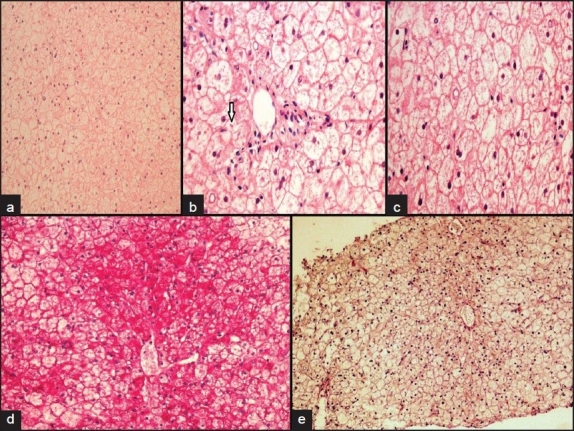
Liver histopathology showed diffuse hepatocyte swelling with rarefaction of cytoplasm and compressed sinusoids (a), intracytoplasmic giant mitochondria seen as round, red to pink globules (arrow) (hematoxylin and eosin stain, ×10) (b), prominent hepatocellular membranes (hematoxylin and eosin stain ×40) (c), abundant cytoplasmic glycogen deposits are demonstrated by a PAS stain ×40 (d), glycogen removed by diastase digestion (10×) (e)