Figure 1.

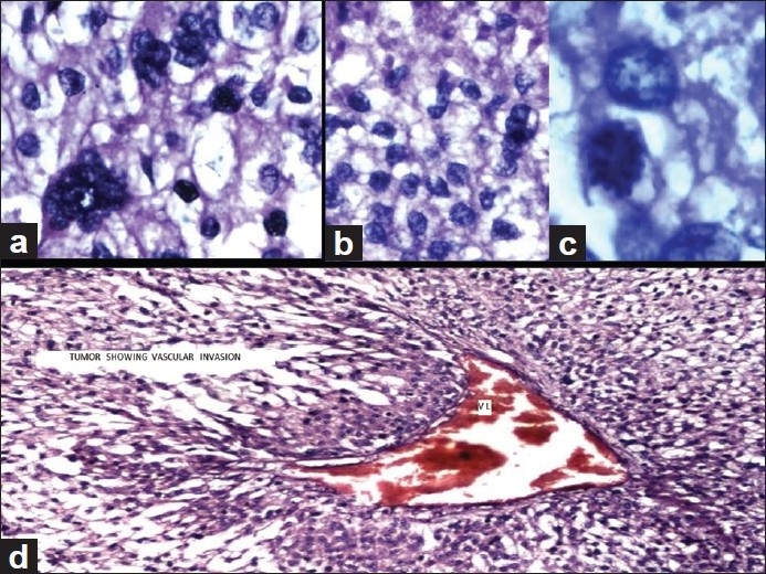
Adrenocortical carcinoma (H and E): (a) Pleomorphic tumor cells showing bizarre hyperchromatic nuclei (×40). (b) Sheet of pleomorphic tumor cells showing necrosis at the top (×40), (c) Mitotic activity by tumor cells (×100), and (d) tumor shows vascular invasion, marked as VI (×20)
