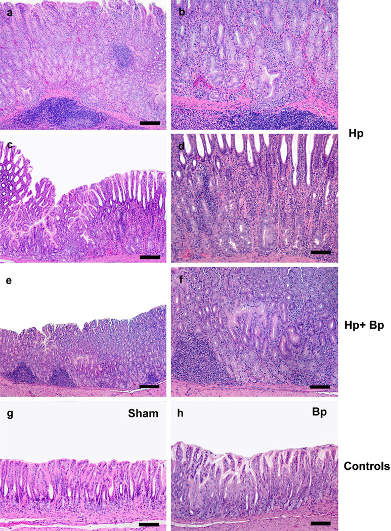
Fig. 1.
Representative H&E images of various groups at 21 WPHPI. Hp monoinfected gerbils had moderate to severe mucosal and submucosal inflammation with occasional distinct lympho-follicular formation (1a, c: 160 µM; b, d: 80 µM), severe glandular and foveolar hyperplasia with prominent villo-papillary epithelial transformation (1a, c) and superficial and basal glandular dysplasia (1, µM and 1c, 160 µM). In HbBp infected gerbils at 21 WPHPI, there was mild to moderate inflammation, mild to moderate glandular hyperplasia and mild glandular dysplasia (1e, 400 µM and 1f, 80 µM). In Sham controls and Bp monoinfected gerbils, the mucosa was mostly within normal limits (1g, h, 80 µM).

