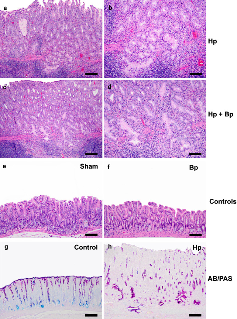
Fig. 2.
Representative H&E images of various groups at 42 WPHPI. Both the Hp only (2a, b: 160 µM) and Hp Bp infected animals (2c, d: 80 µM) exhibited severe mucosal inflammation with lympho-follicular formation, glandular hyperplasia, glandular mucinous and/or globoid cytoplasmic change and moderate to severe glandular dysplasia with frequent herniation of the dysplastic glands into the submucosal lymphoid aggregates. In sham and Bp only infected controls, the mucosal changes were none to minimal. On Alcian blue/PAS staining, pH 2.5, the stomach of Hp infected gerbils at WPHPI showed predominant cytoplasmic gastric type (neutral) red mucin expression within the hyperplastic and metaplastic/dysplastic foci with loss of intestinal acidic mucin expression (blue staining) in basal glands as usually seen in the controls (2 g, h, 160 µM). (For interpretation of the references to color in this figure legend, the reader is referred to the web version of this article).

