Figure 2.
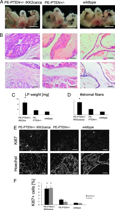
IKK2 activity on PTEN+/- background increases tumor size and epithelial and stromal proliferation. (A) Steromicroscopical images of the structures surrounding the urethra (u), bladder (bl), and seminal vesicles (SV) from 12-month-old mice. The prostate lobes are seen between the noted structures. (B) H&E stain of lateral prostates at 12 months, focusing on epithelium (upper panels) and stroma (lower panels). The genotypes are according to labeling in A. (C) Quantification of dry prostate weight of lateral prostates at 12 months. (D) Stromal fiber number in between epithelial ducts (see Materials and Methods) of 12-month-old lateral prostates. (E) Paraffin sections of lateral prostates (12 months) stained for proliferation marker Ki67. Positive cells in the epithelium (arrowhead) and stroma (arrow) are seen. Hoechst staining of the same sections (lower panels) are given to indicate tissue structure. (F) Quantification of Ki67-positive cells in the epithelium and stroma for the indicated genotypes. Scale bars, 5 mm (A); 50 µm (B, E). Error bars, SEM. *P < .01.

