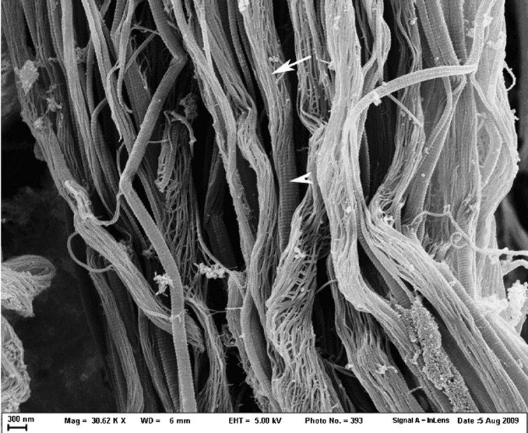
Fig. 3.

Rat tail tendons treated with 25 mM NKISK for 5 days (30 620× magnification). With longer treatment time, dissociation into subfibrils became much more extensive. Intact fibrils can be seen as well as a markedly dissociated fibrils and fibrils in transition from an intact state (arrowhead) to a dissociated state (arrow). Note that the subfibrils appear to be arranged in a right-handed spiral.
