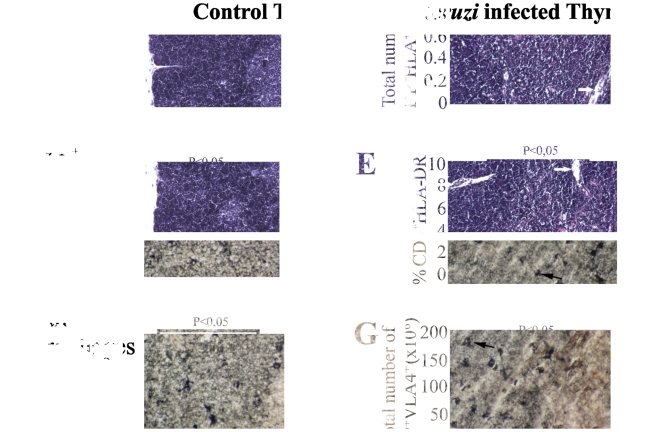
Figure 1. Thymic atrophy in BALB/c acutely infected with T. cruzi.
The histological profiles show the (upper left) normal thymic architecture (C, cortex; M, medulla) and (upper right) marked cortical and medullary atrophy in the thymus of an acutely-infected mouse. (lower left) The numerous metallophilic macrophages are present in the cortico-medullary zone (arrows) of normal thymus. However, following T. cruzi acute infection (lower right) the number and distribution of metallophilic macrophages are changed with the cells dispersed throughout not only the cortico-medullar area but also the cortical region. Infected mice were evaluated herein at day 15 post-infection. These data are representative of two independent experiments using four mice per group.

