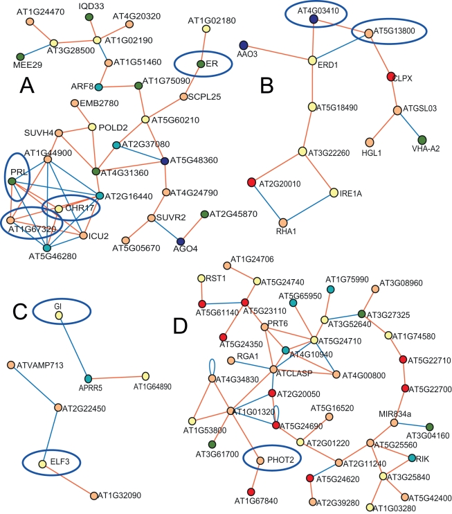
Figure 7. Self-affiliated expression networks of GWA mapping significant candidates.
Shown are the co-expression networks that did not involve any GSL-affiliated genes. These networks contain the ER locus (A), the CLPX locus (B), the ELF3 and GI loci (C), and the PHOT2 locus (D). Triangles show genes known or predicted to be involved in glucosinolate biology, while circles are other genes. White symbols show those genes with no significant GWA in these studies. Other colors show GWA candidates in the listed dataset: Apricot for control; teal for silver; yellow for seed; olivegreen for control and silver; blue for control and seed; cyan for control and seed; red is for all three datasets. Circled in blue are genes that were selected for validation in the current study (Table S9).

