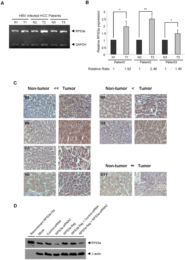
Figure 2. Expression of RPS3a in HBV-associated hepatocellular carcinoma tissues.
(A) Detection of endogenous RPS3a mRNA levels in the tumor and non-tumor regions of HBV-associated HCC tissues. Total RNA was extracted from tumor or non-tumor tissues of three HBV-caused HCC patients. Using total RNA extracts (2 µg) and oligo dT primer, cDNA was synthesized. N and T represent the non-tumor tissues and tumor tissues, respectively. (B) Relative expression ratio of RPS3a mRNA between the tumor and non-tumor tissues of HBV-associated HCC. Data were calculated by three independent experiments (* P<0.05 and ** P<0.001). Data represent the intensity ratio of RPS3a band after normalization to GAPDH in the Bio-1D image analysis software. (C) Representative immunohistochemistry data of endogenous RPS3a protein expression in the HBV-associated HCC tissues. The difference of RPS3a expression between non-tumor and tumor tissues was determined by visual inspection under microscope (magnification ×400) and assigned on the top of the panels. See Table 1 for full information. (D) Generation and validation of human RPS3a antibody. Human RPS3a protein was produced in E. coli and the rabbit antibody was generated. The specificity of the generated antibody was validated by knock-down and over-expression of RPS3a in Huh7 cells. Huh7 cells were harvested at 72 hr after transfection.

