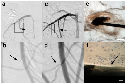
Figure 2. Follow Up of the Detached Distal Tips.
In a. the initial follow up angiogram directly following detachment in the Superior Mesenteric Artery (SMA) is shown with a square marking the blow-up in b. Arrows indicate the detached distal tip. In c. an SMA angiogram, performed 80 days after the intervention in the same animal, is shown with a square indicating the blow-up in d. Arrows indicate the detached distal tip. In e. a microphotograph of a histological van Geeson and toulene blue staining prepared by grind-cutting with a detached tip in situ, is shown. Scale bar = 100 µm. The blow-up in f shows the parylene coating surrounding the detached tip which is marked by an arrow. Note that no fibrous response or inflammation is observable. Scale bar = 4 µm.

