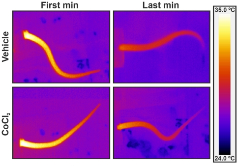
Figure 3. Infrared images of cutaneous temperature.
Tail infrared digital images of representative rats which received either vehicle or CoCl2 into lateral septal area, during the first and last minute of restraint. Note the drop in cutaneous tail temperature during the restraint. All images use the same color coding for temperature.

