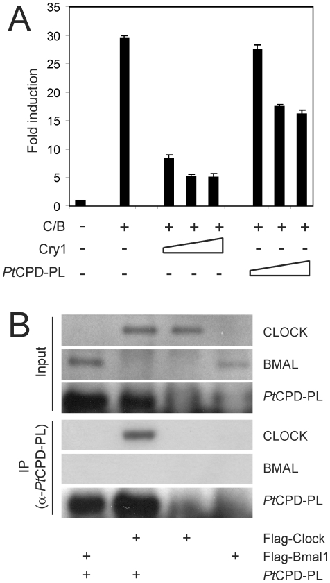
Figure 4. PtCPD photolyase represses CLOCK/BMAL1-driven transcription and interacts with CLOCK.
(A) COS7 cell-based CLOCK/BMAL1 transcription assay using a mPer1 E-box promoter-luciferase reporter construct. Luminescence, shown as x-fold induction from the basal expression level (set to 1), is indicated on the Y axis. pcDNA3, pRL-CMV, and the mPer2::luc were added in all reactions. The presence or absence of Cry1 (10-100 ng) and PtCPD-PL (100-300 ng) expression plasmids is indicated below the graph. Empty pcDNA3 vector was added to correct for the amount of DNA transfected. Mean and standard deviation of triplicate samples are shown. (B) Identification of photolyase-binding proteins. PtCPD-PL was precipitated from HEK293T cells, transfected with PtCPD-PL, Flag-Clock or Flag-Bmal1 or double transfected with PtCPD-PL and either Flag-Clock or Flag-Bmal1. Upper panels: Immunoblot analysis of total cell lysates, confirming the presence of the various transiently expressed proteins. Lower panels (IP): Immunoblot analysis of precipitated PtCPD-PL (anti-PtCPD-PL antibodies) and CLOCK and BMAL1 (anti-FLAG antibodies).

