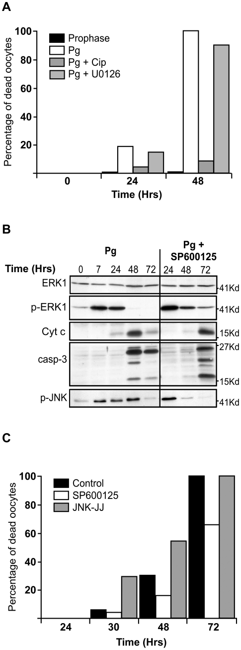
Figure 5. Egg apoptosis involves JNK and Cdk1 activities.
(A) Prophase oocytes were either incubated with a MEK inhibitor (U0126, grey columns) or injected with the p21Cip1 protein (Cip, crosshatched column) and then stimulated (Pg, white column) or not (prophase, black column) with progesterone. Egg death was monitored by following external egg morphology at the indicated times (in hours, Hrs) after progesterone addition (+Pg). (B) Prophase oocytes were stimulated with progesterone (Pg, time 0). Seven hours later, metaphase II-arrested eggs were incubated with or without the JNK inhibitor, SP600125. Egg proteins were analysed at the indicated times by immunoblot with antibodies against ERK1, the active phosphorylated form of ERK1 (p-ERK1), Cyt c, active caspase 3 (casp-3) and the active phosphorylated form of JNK (p-JNK). (C) Oocytes were induced to mature as in (B) and were then incubated with (white column) or without (black column) the JNK inhibitor, SP600125, or injected with mRNA encoding a constitutive active form of JNK (JNK-JJ, crosshatch columns). Egg death was monitored by following external egg morphology for all these three different conditions at the indicated times.

