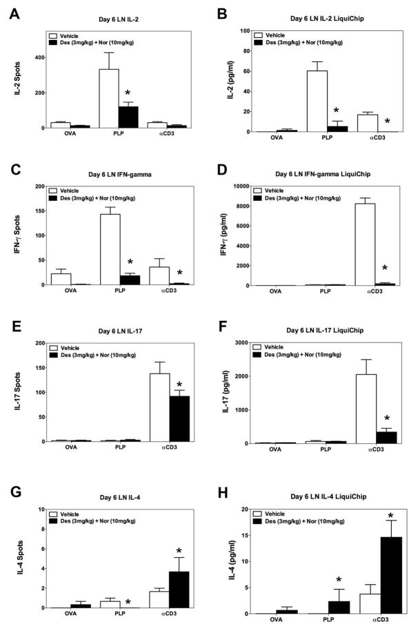
Fig 7. Decrease in the number and level of inflammatory cytokine produced ex vivo by lymph node cells.
SJL/J mice received 3×10⁶ naive PLP139–151-specific TCR transgenic T cells (5B6 T cells) on day -3 prior to PLP139–151 in CFA priming. Groups of 5 mice received either Vehicle, or a combination of desloratadine (3mg/kg) plus nortriptyline (10mg/kg) via gavage on days 0–10 post PLP139–151 sensitization. On day 6 lymph nodes were collected and total lymph node cells (5×10⁵ cells per well) activated ex vivo in the presence of OVA323–339 (20μM), PLP139–151 (20μM), or anti-CD3 (1μg/ml) in ELISPOT plates and culture plates to determine the number of IL-2 (A), IFN-γ (C), IL-17 (E), and IL-4 (G) secreting cells and level IL-2 (B), IFN-γ (D), IL-17 (F), and IL-4 (H) secreted, respectively. The level of cytokine secreted was determined via 10-plex LiquiChip. The data is presented as the mean number of cytokine secreting cells, and level of cytokine secreted in pg/ml from lymph node cells collected from individual mice reactivated in triplicate wells. An asterisk symbol (*) indicates a p value < 0.05 in comparison to Vehicle treated mice. One representative experiment of two is presented.

