
Figure 1.
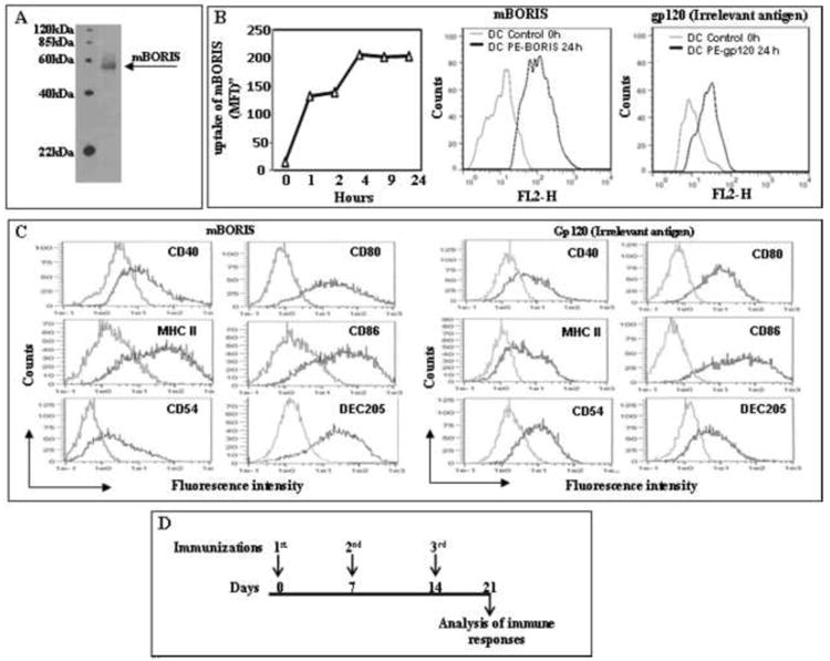
Preparation of DC/mBORIS vaccine and experimental design of immunological studies. (A) Analysis of purified mBORIS recombinant protein in 10% Bis-Tris gel after Coomassie blue staining. (B) The levels of mBORIS proteins (1μM) uptake by CD11c-enriched DC at the indicated time points was analyzed by FACScan using intracellular staining method with polyclonal antibody specific to mBORIS followed by secondary anti-rabbit antibodies. Representative histograms showing uptake of mBORIS and control gp120 proteins after 24 hours are presented. (C) Flow cytometric analyses of DC cell surface phenotype. Cells were incubated in the presence of GM-CSF/IL-4 (see Materials and Methods) and loaded with mBORIS and gp120, respectively, followed by detection of CD40, CD80, CD86, MHC class II, CD54, and DEC205 molecules (black line) or isotype control primary Abs (gray line) in the CD11c-enriched cell population. Representative histograms from 2 experiments are presented. (D) Immunization schedule for administration of DC-based mBORIS and gp120 vaccines.