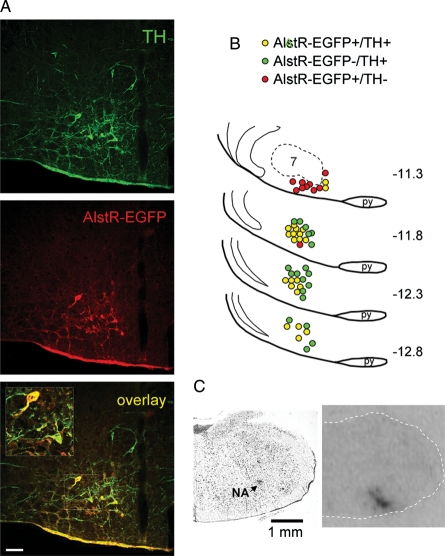
Figure 1.
Targeting catecholaminergic C1 neurones in the rostral ventrolateral medulla oblongata (RVLM) with PRSx8-AlstR-EGFP-LV (A) Confocal images of tyrosine hydroxylase (TH)-positive neurones (green) expressing AlstR-EGFP (red). Bregma level -12.8 mm. Scale bar = 50 µm. (B) Distribution of TH-immunoreactive neurones expressing AlstR-EGFP in one representative rat brainstem 5 weeks after microinjection of PRSx8-AlstR-EGFP-LV into the RVLM. Each symbol represents three neurones. Numbers indicate distances from Bregma. 7, facial motor nucleus; py, pyramidal tract. (C) Binding of 125I-allatostatin in the rat RVLM 9 days after microinjection of PRSx8-AlstR-EGFP-LV. Left, Nissl stain; right, autoradiography image showing strong AlstR binding in the rostral ventrolateral reticular formation. NA, nucleus ambiguus.

