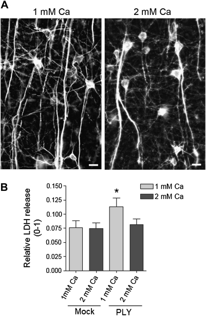
Figure 4.
A, Verification of the MAP2 immunostaining of pyramidal neurons in the cortex of mice 8 hours after preparation of acute slices in ACSF with both 1 and 2 mM Ca shows completely intact neurite morphologies. Scale bars: 20 μm. B, LDH release from brain slices, incubated for 4 hours with or without 0.2 μg/mL PLY in the presence of 1 and 2 mM extracellular Ca. * P < .05 vs all, Mann–Whitney U test (see Methods). Values represent mean ± SEM, n = 6 experiments (5 slices/condition/experiment). ACSF, artificial cerebrospinal fluid.

