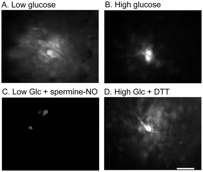
Fig. 6. Lucifer yellow labeling in astrocyte cultures grown in low and high glucose.
Astrocytes were grown in culture medium containing low (5.5 mmol/L, A, C) or high (25 mmol/L, B, D) glucose for two weeks. Single astrocytes were impaled with a micropipette containing 4% Lucifer yellow, and the dye was allowed to diffuse for 2 min, and dye-labeled area measured (see Table 2). (C) Astrocytes grown in low glucose were treated with spermine-NO (250 μmol/L for 1h) prior to dye transfer assay. (D) Astrocytes grown in high glucose were treated with dithiothreitol (DTT, 10 mmol/L for 10 min) prior to dye transfer assay. The scale bar represents 40 μm and applies to all panels.

