
Figure 1.
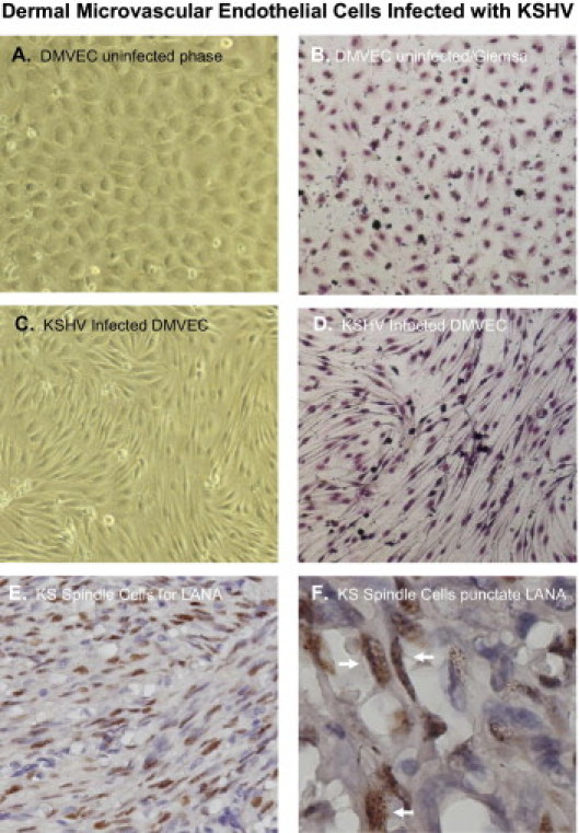
Human dermal microvascular endothelial cell (DMVEC) mock-infected controls and DMVEC cells infected with KSHV for 10 days. For infections, cells were treated with BCBL-1 virus at a multiplicity of infection (moi) of 0.01 and cultivated in EBM-2 complete medium. Phase images were taken with a NIKON TE 2000S microscope mounted with a CCD camera. Shown are phase (A) and Giemsa (B) stained images of mock-infected cells showing a cobblestone-like morphology C: KSHV-infected cells with the characteristic spindle shape morphology. D: This type of swirling/spindling appears more pronounced when a Giemsa stain is performed and appears very distinct from uninfected cells. E: An IHC stain of a nodular AIDS-KS tumor showing KSHV LANA positive spindle cells with a hematoxylin counterstain. F: Shows KS tissue with spindle cells stained positive for KSHV LANA with a nuclear punctate staining pattern (white arrows). All images were, with the exception of Figure 1F, taken at a total magnification of ×200. The image in Figure 1F was taken at a total magnification of ×600 to discern the nuclear punctate staining pattern of KSHV LANA.