
Figure 10.
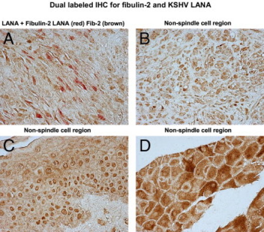
Fibulin-2 expression in human KS tumor tissue. Briefly, paraffin-embedded KS tissue was deparaffinized in xylene and hydrated in graded alcohol, and antigen unmasking was performed in citrate buffer in a microwave. Cells were stained by dual-labeled IHC for KSHV LANA (Vector Red, red) and fibulin-2 (DAB brown). No counterstain was used in this procedure. Bright-field images were photographed on a Nikon TE2000S microscope at total magnification of ×200. A: A spindle cell region; B–D: non-spindle cell regions observed in a representative KS tissue specimen.