
Figure 2.
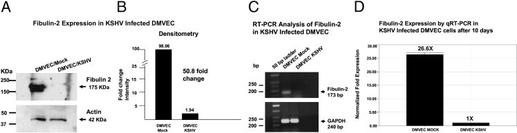
Analyses for fibulin-2 expression at 10 days after mock or KSHV infection of DMVEC. A: Lysates (15 μg) from mock- and KSHV-infected DMVEC were separated by electrophoresis in 4% to 20% PAGE gels transferred to nitrocellulose membranes and screened for expression of fibulin-2 using a rabbit polyclonal antibody (Santa Cruz Biotechnology) at a 1:2,000 dilution. Western blot bands show evidence of a ∼175 kDa protein in lysates from mock-infected DMVEC compared to the mark reduction of this band in KSHV-infected DMVEC. B: Densitometry analysis of the Western blots in Figure 2A reveals a greater than 50-fold reduction in fibulin-2 protein expression in KSHV-infected DMVEC cells at 10 days compared to mock-infected control cells. C: Semiquantitative RT-PCR analysis of fibulin-2 mRNA expression in KSHV-infected DMVEC. Ten nanograms of cDNA from mock- and KSHV-infected DMVEC were amplified by PCR using fibulin-2 gene–specific primers. PCR products were electrophoresed in 1.5% agarose, and PCR DNA fragments were sized using a 50-bp ladder. The expected fragment size for fibulin-2 is 173 bp. GAPDH was amplified as a loading control with a fragment size of 240 bp. D: Real-time RT-PCR analysis of fibulin-2 expression in KSHV-infected DMVEC cells 10 days after infection compared to mock-infected control cells. Relative fold expression was normalized to GAPDH.