
Figure 2.
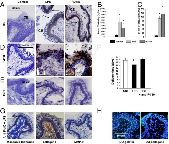
Complement activation and macrophages/monocytes infiltration in the cervix of LPS- and RU486-treated mice. A: Immunohistochemical determination of C3 in cervical tissue. Arrows indicate C3 deposition (brown staining) in the columnar epithelium (CE) in LPS- (middle panel) and RU486-treated mice (right panel). No complement deposition was observed in the cervical tissue of the control mice (left panel). Original magnification is ×20. B: Serum C3adesArglevels in control, LPS-, and RU486-treated mice. Statistically different from control, *P < 0.05 (n = 5 to 6 mice/group). C: Serum C5adesArglevels in control, LPS-, and RU486-treated mice. Statistically different from control, *P < 0.05 (n = 5–6 mice/group). D: F4/80 staining in cervical tissue. Increased macrophage infiltration (brown color) was observed in the cervical stroma, epithelium, and mucus of LPS- (middle panel) and RU486-treated mice (right panel). Few macrophages were observed in the cervix of age-matched control mice (left panel). Original magnification is ×20. The inserts in the middle and right panels include macrophages shown at a higher magnification. E: Gr-1 staining in cervical tissue. Few Gr-1+ neutrophils cells were found in the cervical stroma of control (left panel), LPS- (middle panel), and RU486-treated mice (right panel). Original magnification is ×20. F: Delivery time in control, LPS-, and LPS plus anti-F4/80 (n = 4–6 mice/group). Significantly different from control, *P < 0.05. G: Trichrome (TC) staining for collagen I and for matrix metalloproteinase (MMP)-9 in cervical tissue of LPS-treated mice depleted of macrophages with anti-F4/80. Abundant collagen fibers were shown with TC staining (left panel) and by IHC with antibodies against collagen I (middle panel). Staining for MMP-9 was negative in F480 plus LPS-treated mice (right panel). H:In situ zymography using DQ-gelatin and DQ-collagen I as substrate in LPS-treated mice pretreated with anti-F4/80. Gelatin and collagen I degradation was not observed in cervical tissue from F4/80 plus LPS-treated mice. Original magnification is ×20.