
Figure 3.
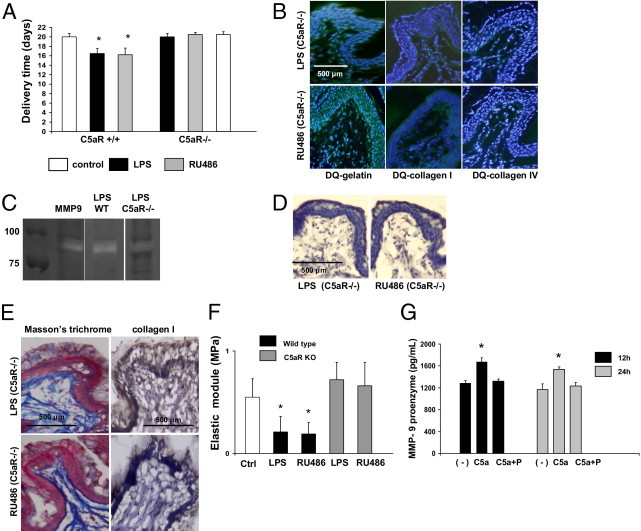
C5aR deficiency and progesterone prevents preterm labor/delivery (PTD). A: Delivery time in control and LPS- or RU486-treated, wild-type, C5aR+/+ and C5aR−/− mice (n = 5 to 7 mice/group). Significantly different from control, *P < 0.05. B:In situ zymography using DQ gelatin (left panels), DQ collagen I (middle panels), and DQ collagen IV (right panels). In contrast to the increased fluorescence (increased gelatin and collagen degradation) observed in the cervix of LPS- (Figure 1E, middle panels) and RU486-, wild-type treated mice (Figure 1E, right panels), gelatin and collagen degradation was not observed in LPS- (upper panels) and RU486-treated, C5aR−/− mice (lower panels) comparable to control cervical tissue (Figure 1E, left panels). C: Gel zymography of nonreduced cervical tissue lysates. Purified matrix metalloproteinase (MMP)-9 was used as a control. Increased MMP-9 was observed in wild-type (WT) mice treated with LPS. D: Immunohistochemical detection of MMP-9 revealed weak staining for MMP-9 in LPS- (left panel) and RU486-treated, C5aR−/− mice (right panel). E: Collagen detection by trichrome staining and IHC. Dense network of collagen fibers was observed in the cervix of LPS- (upper panels) and RU486-treated (lower panels) C5aR−/− mice in contrast with the disperse thin fibers of collagen observed in the cervix of LPS- and RU486-treated, wild-type, C5aR+/+ mice (Figure 1D, middle and right panels, respectively). F: Biomechanical studies of the cervix. The elastic modulus (measure of the tissue stiffness) was decreased in the cervical tissue of LPS- and RU486-, wild-type treated mice when compared to control mice. Decreased elastic module was not observed in LPS- and RU486-treated C5aR−/− mice, indicating an increase in the stiffness of the cervix in these mice. Elastic module values in LPS- and RU486-treated mice were comparable to age-matched control mice (n = 4 mice/group). Significantly different from control mice (Ctrl), *P < 0.05. G: Complement activation triggers release of pro-MMP-9 by murine macrophages in vitro. Splenic macrophages were stimulated with C5a and C5a plus progesterone (P). Culture supernatants were collected after 12 and 24 hours and analyzed for MMP-9 by enzyme-linked immunosorbent assay (n = 4 experiments/group; *P < 0.05 versus untreated cells).