
Figure 5.
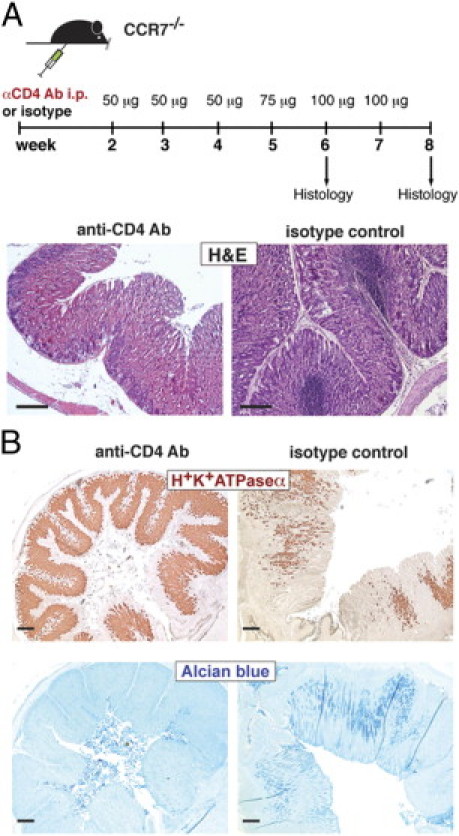
CD4+ T cells are crucial for the development of AIG and ectopic follicular aggregates. A: Schematic representation of CD4+ T-cell depletion in CCR7−/− mice. CCR7−/− mice were treated with 50 to 100 μg (as indicated) of anti-CD4 antibody or isotype control antibody i.p. starting at 2 weeks of age up to 6 or 8 weeks of age (once per week). B: Paraffin-embedded stomach sections of anti-CD4 Ab- (n = 5) or isotype control antibody-treated mice (n = 7) were stained with H&E for parietal cells (brown; H+/K+-ATPase α; upper panel) or acidic mucopolysaccharides (Alcian blue; lower panel). Scale bar = 200 μm.