
Figure 3.
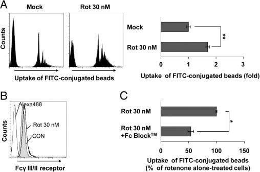
Rotenone (Rot) exposure triggers phagocytic activity in microglia. A: BV2 microglia were cultured in the presence or absence of 30 nmol/L Rot for 24 hours, and phagocytosis of FITC-conjugated fluorescent beads was determined by flow cytometric analysis, as described in Materials and Methods. **P < 0.005 when compared with mock-treated cells. B: BV2 microglia were treated with 30 nmol/L Rot for 24 hours, after which expression of Fcγ receptors was determined by FACS analysis using an anti-CD16/CD32 antibody directed against FcγIII/II receptors. C: BV2 microglia were incubated with 10 μg/mL Fc Block (BD Biosciences) for 1 hour at 37°C and followed by exposure to 30 nmol/L Rot for 18 hours. The phagocytic activity was analyzed by FACS using FITC-conjugated fluorescent beads. *P < 0.05 when compared with Rot alone–treated samples. CON indicates control.