
Figure 6.
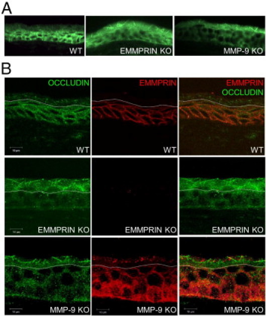
EMMPRIN and occludin expression in corneal epithelium of wild-type, EMMPRIN, and MMP-9 KO mice. A: Immunohistochemical analysis of occludin (green) expression revealed different pattern of expression between the WT and the KO corneal epithelium. In both EMMPRIN and MMP-9 KO, stronger apical occludin membrane staining appearing as longer and thicker lines than in the WT epithelium. B: Double labeled confocal immunohistochemistry of EMMPRIN and occludin in WT and KO mice. In WT mice, EMMPRIN (red) is mainly expressed in the basal layers, gradually decreasing toward the apical layers where its expression is barely detected. The typical membranous staining of occludin (green) is only observed in the apical areas where EMMPRIN staining is absent. Occludin membranous staining is much more pronounced in both EMMPRIN and MMP-9 KO mice and appears in deeper apical layers of the epithelium as longer and thicker lines. Note that the white dotted line was drawn at the point where occludin staining becomes membranous. Below this line occludin staining appears to be mainly cytoplasmic. In WT corneas, this line also corresponds to the extinction of EMMPRIN staining. Above this line, the expression of EMMPRIN occasional detected as is shown in the MMP-9 KO panel, was dissociated from the membranous occludin staining.