
Figure 4.
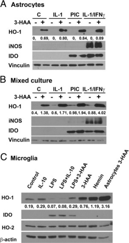
HO-1 expression in human fetal CNS cell cultures. Cultures of astrocytes (A), mixed neurons and glia (B), and microglia (C) were prepared as described in Materials and Methods and treated with 3-HAA (100 μmol/L), recombinant cytokines (all 10 ng/mL), or TLR ligands for 24 hours as described in the Figure 2 legend. Hemin (10 μmol/L) was included as a known inducer of HO-1. Western blotting was performed using a polyclonal rabbit IgG against HO-1 (from Abcam, originally from Stressgen). The blots were stripped and reprobed for iNOS, IDO, HO-2, vinculin, or β-actin (controls for protein loading). The numbers are densitometric ratios of HO-1 to vinculin (A and B) or β-actin (C). The results together indicate that 3-HAA is a strong stimulus for HO-1 induction in human astrocytes and that proinflammatory cytokines and TLR ligands synergize with 3-HAA in the induction of HO-1. However, microglial HO-1 expression was much lower and was regulated differentially by IL-10 and TLR ligands (LPS). The results are representative of five independent experiments for A and B and two for C, with identical results.