
Figure 5.
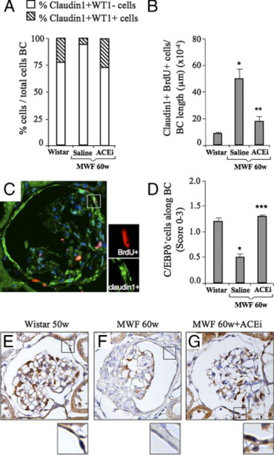
ACE inhibitor reduces proliferation of Bowman's capsule cells and regulates the expression of the cell cycle inhibitor C/EBPδ. A: In the Bowman's capsule of 60-week-old MWF rats given saline, the percentage of claudin1+WT1− cells (white bars) was markedly enhanced, and claudin1+WT1+ cells (striped bars) decreased with respect to Wistar rats. ACEi restored the percentage of both cell populations to control levels. B: Cell proliferation of claudin1+ cells along the Bowman's capsule was enhanced in 60-week-old MWF rats given saline as compared with controls. ACEi treatment significantly limited the number of proliferating cells within the Bowman's capsule (claudin1+BrdU+). *P < 0.05 versus Wistar; **P < 0.01 versus 60w MWF + saline. C: Representative picture of immunofluorescence staining for claudin1 (green) and BrdU (red) in the Bowman's capsule of 60-week-old MWF rats. Inset shows a cell expressing both markers. Nuclei are stained with DAPI (blue). Original magnification, ×630. D: The expression of C/EBPδ in the Bowman's capsule, evaluated by semiquantitative score, decreased at 60 weeks of age in MWF rats receiving saline as compared with Wistar rats, and was normalized by ACEi treatment. *P < 0.05 versus Wistar; ***P < 0.05 versus 60w MWF + saline. Representative photomicrographs of C/EBPδ expression in Wistar (E) and MWF rats given saline (F) or treated with ACEi (G) are shown. Note that in ACEi-treated rats, as in Wistar rats, most of the cells were positive for C/EBPδ (brown signal at high magnification); in MWF rats, the majority of the cells did not express the marker. Nuclei (blue) were counterstained with hematoxylin. Original magnification, ×400.