
Figure 3.
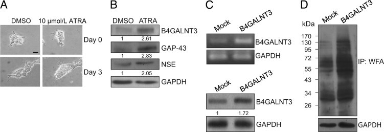
B4GALNT3 expression in SK-N-SH NB cells. A: ATRA-induced SK-N-SH cell differentiation and B4GALNT3 expression. SK-N-SH cells were treated with 10 μmol/L ATRA for 3 days. Dimethyl sulfoxide (0.1%) (DMSO) treatment was used as a control. The differentiation of SK-N-SH cells was shown by increased neurite outgrowth. Scale bar = 10 μm. B: Expression of B4GALNT3 was increased in ATRA-treated SK-N-SH cells as demonstrated by Western blot analysis. The neuronal differentiation induced by ATRA was confirmed by increased expression of GAP-43 and neuron-specific enolase (NSE). Glyceraldehyde-3-phosphate dehydrogenase (GAPDH) is an internal control. The relative band signals were quantified by ImageQuant software version 5.1 as shown. C: Forced expression of B4GALNT3 in SK-N-SH cells. SK-N-SH cells were transfected with pcDNA3.1 (mock) or pcDNA3.1/B4GALNT3 and were selected with G418 for 10 days. The G418-resistant cells were pooled, and reexpression of B4GALNT3 mRNA and B4GALNT3 protein expression were demonstrated by RT-PCR (top panel) and Western blot analysis (bottom panel), respectively. GAPDH is a loading control. D: B4GALNT3 reexpression increases the expression of LacdiNAc structure. The expression of LacdiNAc was detected by Western blot analysis with WFA. GAPDH is a loading control. The molecular weight marker is shown on the left. IP, immunoprecipitated.