
Figure 2.
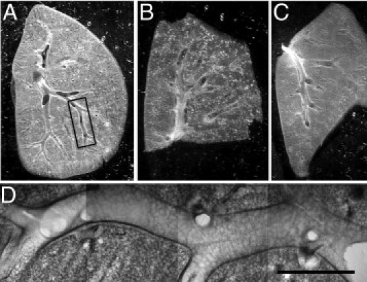
Lung section preparations suitable for dynamic imaging of airway mucosal DCs. A-C: Low-magnification photomicrographs of freshly prepared sections made from the superior lobe (A) and the inferior lobe (B) of the right lung and from the left lung (C). The sections were cut in the level of the main conducting airways. D: Typical location used for two-photon microscopy. Mosaic of four microscopic images obtained with a ×10 objective lens corresponding to the boxed region shown in A. Scale bar = 1 mm.