
Figure 4.
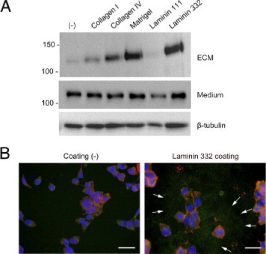
Deposition of Ecto-ColXVII in the ECM is enhanced by specific ligands. A: Immunoblot of the ECM and culture medium. The top panel shows that collagen XVII expressing 293 cells cultured on uncoated plastic (−) deposited negligible amounts of the Ecto-ColXVII in the ECM. The deposition was significantly increased when the cells were cultured on collagen IV, Matrigel, and laminin 332 but not on laminin 111. The Ab HK139 was used to detect the shed Ecto-ColXVII in the ECM. The middle panel shows the shed Ecto-ColXVII in culture medium, as detected by the Ab NC16A-3. The bottom panel shows β-tubulin in cell lysates. B: Consistent with the previous data on immunoblotting, IIF with the Ab HK139 detected shed Ecto-ColXVII (green; arrows) around cells cultured on laminin 332 coating. Red, rhodamine phalloidin; blue, DAPI. Scale bars = 40 μm.