
Figure 2.
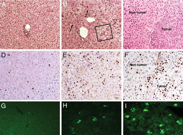
Age-related hepatocellular proliferation with abatement of hepatic steatosis in ACOX1−/− mice. H&E (A–C), BrdUrd (D–F), and terminal deoxynucleotidyl transferase-mediated dUTP nick-end labeling (G–I) staining of liver from 2-week-old (A, D, and G), 5-month-old (B, E, and H), and 1-year-old (C, F, and I) ACOX1−/− mice. Boxed area in B indicates microvesicular steatosis; arrows point to regenerated hepatocytes. In C and F, tumor and nontumor areas are shown.