
Figure 3.
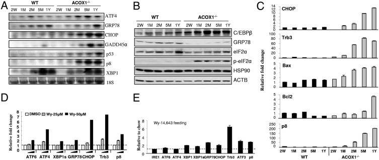
Spontaneous PPARα activation in ACOX1−/− mouse liver aggravates ER stress. A: Northern blot analysis of wild-type (WT) and ACOX1−/− mouse liver RNA. Mice 2 weeks old (W), 1, 2, and 5 months old (M), and 1 year old (Y). B: Western blot analysis of liver protein (20 μg protein). ACTB served as loading control. C: qPCR analysis of liver RNA from wild-type (WT) and ACOX1−/− mice. D: qPCR analysis of ER stress gene expression in primary hepatocytes treated with Wy-14,643. E: ER stress response gene expression in the liver of wild-type mice fed Wy-14,643 for 4 days was analyzed by qPCR. Fold change presented represents normalization of Wy-14,643 group against chow control. 18S was used as internal control.