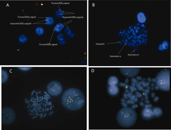
Figure 2. Fluorescent in situ hybridization (FISH) using breakapart probe set for IER3 in MDS samples.
A, Metaphase preparation from patient with t(6;9)(p23.1;q34) demonstrating separation of red signal and green signal. Only a single fusion signal is present from the normal chromosome 6. B, Interphase cells from the same patient. C and D, two other MDS patients in whom IER3 signal was amplified, indicating increased copy number.

