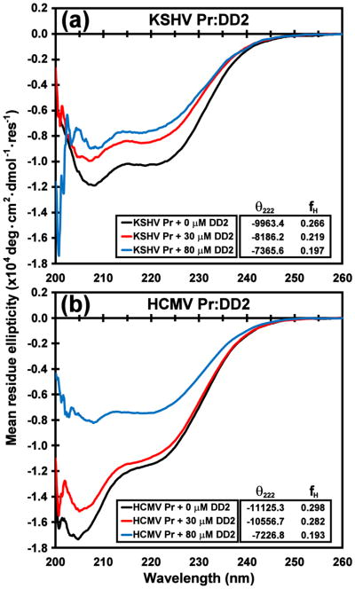
Fig. 8. CD spectra of DD2 titrations with KSHV Pr and HCMV Pr.
The circular dichroism spectra of ~ 3 μM (a) KSHV Pr and (b) HCMV Pr in the presence of 0 μM (black), 30 μM (red), and 80 μM (blue) DD2. Estimated fractional helicity (fH) values derived from the mean residue ellipticity of the 222 nm band are listed in the insets, and indicate loss of helical content with increasing molar equivalents of DD2. Loss of helicity is a strong indication of HHV protease dimer disruption.

