
Figure 4.
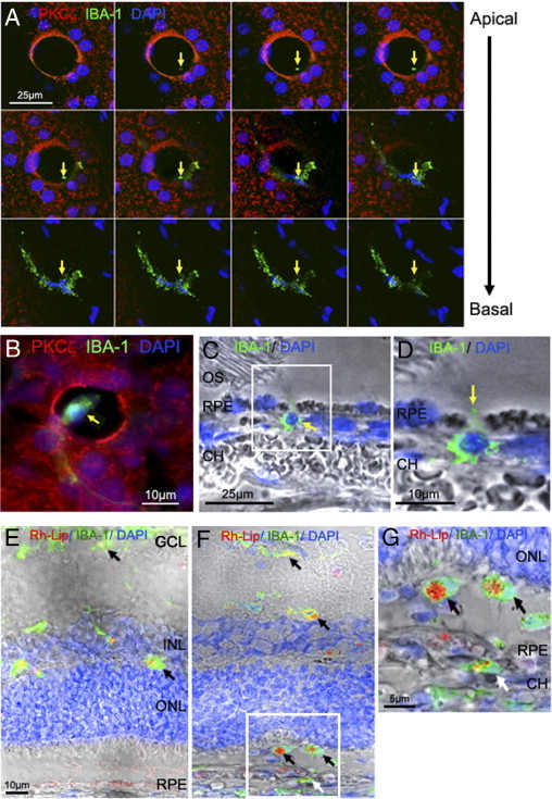
Transcellular migration of microglia/macrophages through RPE pores. A: RPE flat mount from 5-month-old GK rat immunostained with PKCζ (red), IBA-1 (green), and DAPI (blue) and imaged by confocal microscopy. Top view projection of all z-series sections of a representative IBA-1–positive cell (green) in a transcellular pore delimited by PKCζ expression (red). The first Z-sections of the confocal stack analysis highlighted the beginning of the cell process as a green point (arrows). The end Z-sections clearly show a microglia/macrophage within the pore. Scale bar = 25 μm. B: Triple staining of RPE flat mount from 5-month-old diabetic rats by PKCζ (red), IBA-1 (green), and DAPI (blue) show a microglia/macrophage within the pore (arrow). Scale bar: 10 μm. C and D: Sections from 5-month-old GK rat retina immunostained with IBA-1 antibody (green) and DAPI (blue) associated with phase contrast. IBA-1 staining shows a macrophage/microglia passing through the RPE (arrow) (C) and a cytoplasmic extension between 2 RPE cells (arrow) (D). Scale bar: 25 μm (C); 10 μm (D). E–G: Retinal sections 72 hours after intravitreous injection of rhodamine-liposome (Rh-Lip) (red), macrophages/microglia stained by IBA-1 (green). In 5-month-old control rats (E), liposomes are engulfed by IBA-1–positive cells in inner retina (black arrow). At the same age of diabetes in GK rats (F), numerous IBA-1–positive cells having engulfed Rh-Lip are located in the outer retina and subretinal space (black arrows) and under the RPE, in the choroid (white arrow). Higher magnification confirmed that IBA-1–positive cells loaded with Rh-Lip have migrated from the vitreous through the retina and RPE toward the choroid (G). Scale bar: 10 μm (E and F); 5 μm (G). CH = choroid; OS = outer segment of photoreceptor; RPE = retinal pigmented epithelium.