
Figure 7.
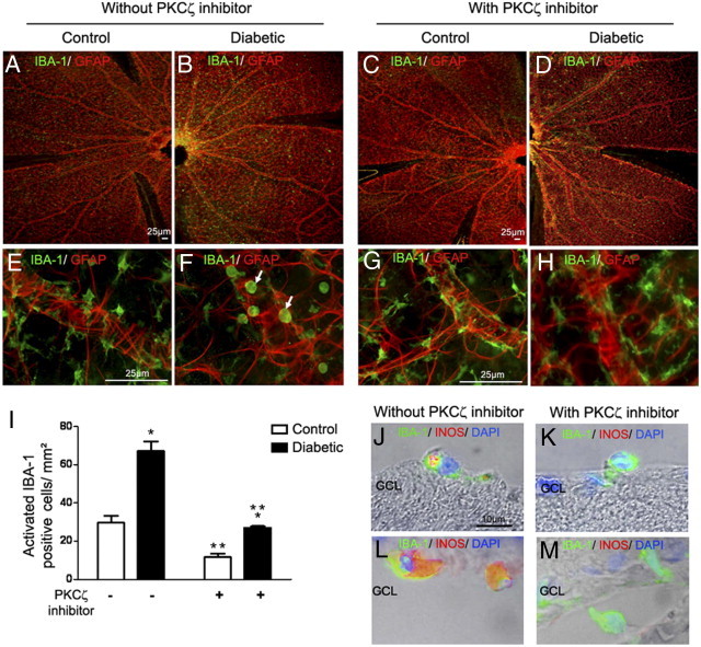
Effects of PKCζ on microglia/macrophages activation.
(A–H) IBA-1 (green) and GFAP (red) immunostaining on whole neuroretina flat mounts from 12-month-old control (A, C, E, and G) or diabetic GK rats (B, D, F, and H). Controls treated with the PKCζ inhibitor (C and G) or not treated (A and E) showed in the retina, microglia with a resting dendritic shape with long branching processes form. By contrast, in diabetic GK rats not treated with PKCζ inhibitor (B and F), microglia showed a round, amoeboid shape (F, arrow), whereas PKCζ inhibition (D and H) induced a change in IBA-1–positive cells morphology, indicating a return to a normal resting microglia (H). Scale bar: 25 μm (A–H). I: Quantification of round activated IBA-1 microglia/macrophages in neuroretina flat mounts. Graph shows the mean number of cells/mm2 counted in four areas in three separate flat mounts. *P < 0.05 versus control; **P < 0.05 versus without PKCζi injection. J–M: Retinal sections from 12-month-old diabetic rats treated or not treated with PKCζ inhibitor. Double staining with IBA-1 antibody (green) and iNOS antibody (red) associated with phase contrast showed that, in diabetic GK rats treated with PKCζ inhibitor (K and M), IBA-1–positive cells do not express iNOS as compared with activated IBA-1–positive cells in diabetic GK rats not treated (J and L). Scale bar = 10 μm. GCL = ganglion cell layer.