
Figure 1.
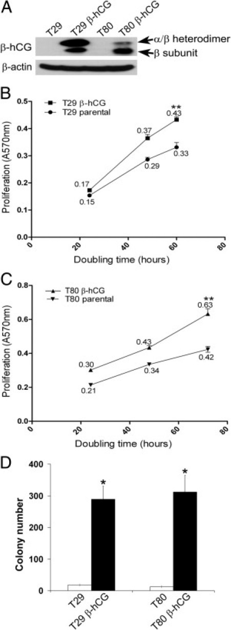
Expression of β-hCG in immortalized ovarian surface epithelial cells promotes cell proliferation and anchorage-independent growth. A: Western analysis revealed elevated expression of the β-hCG subunit alone, as well as the β-subunit in heterodimeric complex with the α-subunit, after β-hCG cDNA infection of immortalized ovarian surface epithelial T29 and T80 cells. B: T29 β-hCG showed a statistically significant increase in the rate of cell proliferation over the course of 24, 48, and 60 hours (slope = 0.0073 ± 0.00044 AU/hour), relative to similar-passage parental T29 (slope = 0.005 ± 0.00046 AU/hour). Numeric values indicate proliferation rate. AU, arbitrary units. **P < 0.0005. C: T80 β-hCG cells similarly showed a statistically significant increase at 24, 48, and 72 hours (slope = 0.0069 ± 0.00057 AU/hour), relative to similar-passage parental T80 cells (slope = 0.0044 ± 0.00057 AU/hour). Numeric values indicate proliferation rate. **P < 0.0005. D: T29 β-hCG and T80 β-hCG cells showed a statistically significant increase in anchorage-independent colony formation, compared with similar-passage parental T29 and T80 cells. Error bars indicate mean ± SD. *P ≤ 0.01; n = 3.