Figure 2.
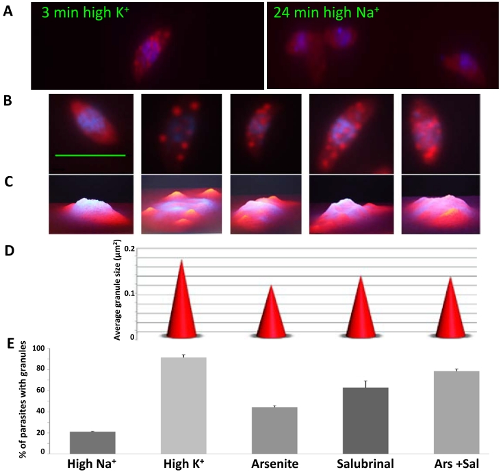
Different mRNA granule inducing conditions generate granules with different characteristics. A) Time course of granule formation. Infected host cell monolayers were scraped in either high K+ buffer or high Na+ buffer, and incubated for different times. Parasites were fixed every 3 minutes (0 to 24 min) and RNA FISH was carried out against total polyadenylated RNA (Cy5, red) followed by DAPI staining (blue). Representative images of times 3 and 24 min in high K+ and high Na+ respectively. B) Representative images of granules formed in different inducing conditions: high Na+, high K+, Arsenite, Salubrinal and Arsenite + Salubrinal (from left to right). C) 3D profile of images shown in B. D) Granule size in different inducing conditions (shown in B). We show the average granule size of 100 parasites. All differences are significant (t tests with p<0.05), except for the comparison Salubrinal with Arsenite + Salubrinal which is not significant. E) Percentage of parasites that contain granules, in different inducing conditions. We counted 200 parasites in triplicate experiments. All differences are significant (t tests with p<0.05), except for the comparison Arsenite with Salubrinal and the comparison Salubrinal with Arsenite + Salubrinal. Scale bar 5 µm.

