
Figure 8.
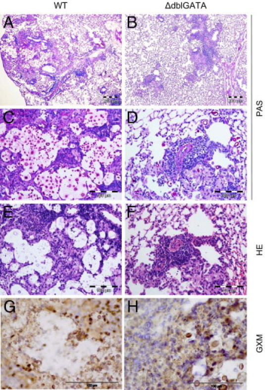
Lung sections from infected WT and ΔdblGATA mice indicate better fungal control in the absence of eosinophils at 60 dpi. A through D: PAS staining. Scale bars: 200 μm (A and B); 100 μm (C and D). E and F: H&E staining. Scale bar = 100 μm. G and H: IHC was performed on sections from the same mice, and glucuronoxylomannan (GXM)–containing foci are brown. Scale bar = 100 μm. ΔdblGATA mice show reduced numbers of cryptococci in the lungs (A–F) and formation of smaller foci of accumulating cryptococci and GXM compared with WT mice (G and H). There was pronounced influx of inflammatory cells in WT compared with ΔdblGATA mice.