
Figure 6.
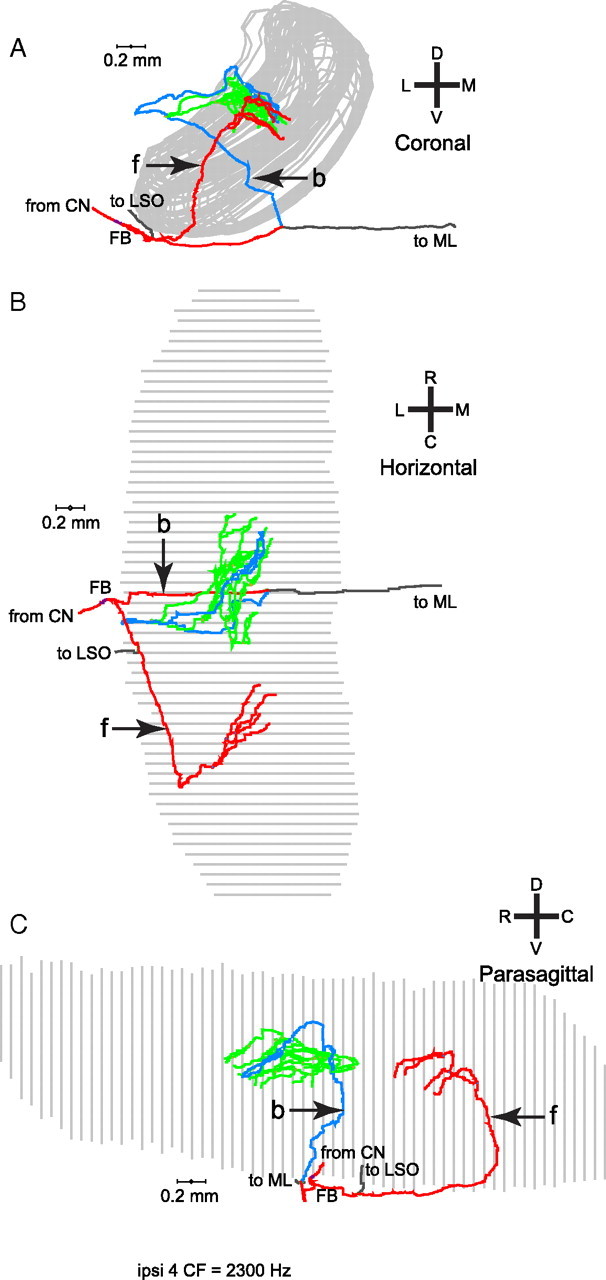
Another example of an ipsilateral MSO projections of one fiber. A, This fiber ran underneath the MSO (coronal view) and also formed forward (f) and backward (b) projecting branches. B, C, The horizontal (B) and parasagittal (C) views reveal that the branches innervate different rostrocaudal portions of the MSO. The backward branches (green and blue) innervated a more rostral portion of the MSO and covered more length of axon from FB than the forward branches.