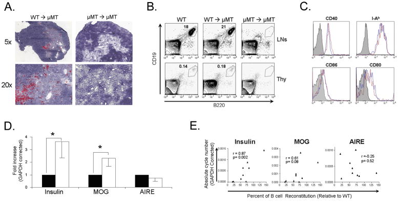
Figure 2.
WT BM restores B cells and thymic insulin and MOG expression in B cell deficient μMT mice. (A) Immunohistochemistry staining for B220 (red) in the thymi of μMT mice receiving either WT BM or μMT BM two months following BM transfer. (B) FACS analysis of CD19+B220+ cells in LNs and thymi of BM recipient mice two months following BM transfer. (C) FACS analysis of B cells in thymi of WT and B cell restored μMT mice two months following BM transfer. Shaded histogram- isotype control, blue histogram- WT, red histogram- B cell restored μMT mice. (D) Real-time RT-PCR analysis of insulin, MOG, and AIRE in thymi of μMT receipt mice transplanted with μMT (n=5) or WT (n=7) BM two months following BM transfer are expressed relative to μMT BM recipients. Solid bars- μMT BM; open bars- WT BM. * p<0.05. (E) Correlation analysis between degree of B cell reconstitution and insulin, MOG, and AIRE expression in thymi of μMT mice receiving WT BM.

