
Figure 4.
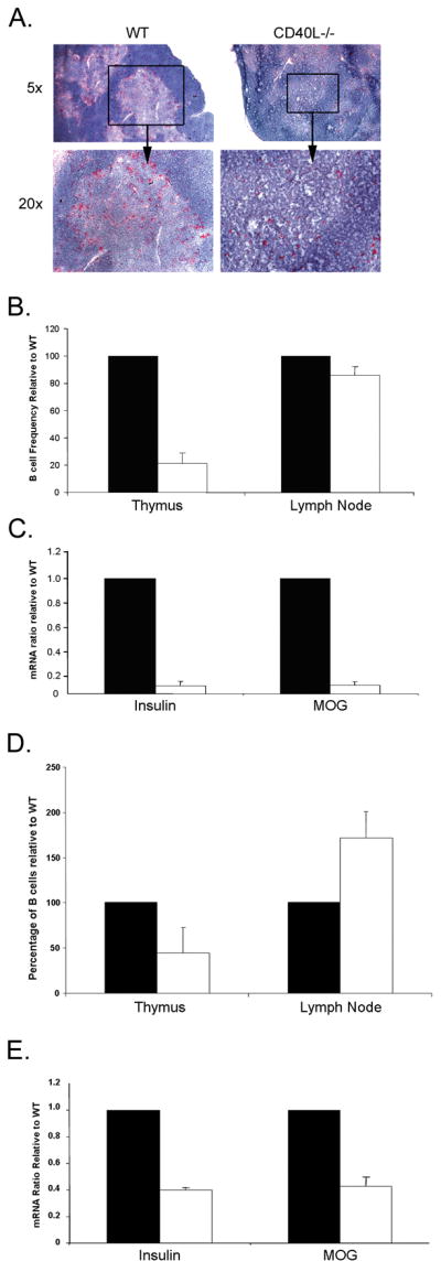
Reduced B cell numbers in CD40L and CCR7 deficient mice is associated with TRA expression. (A) Immunohistochemistry staining for B220 (red) in the thymi of WT and CD40L−/− mice. (B) Frequencies of B220+CD19+ cells as obtained by FACS analysis of LNs and thymi from 4–6 week old WT and CD40L−/− mice. Solid bars-WT; open bars- CD40L−/−. (C) Real-time PCR analysis of TRA and AIRE in thymi of WT (n=3) and CD40L−/− (n=3) mice. Results are expressed relative to WT mice. Solid bars-WT; open bars- CD40L−/−. (D) Frequencies of B220+CD19+ cells as obtained by FACS analysis of LNs and thymi from 4–6 week old WT and CCR7−/− mice. Solid bars-WT; open bars- CCR7−/−. (E) Real-time PCR analysis of TRA and AIRE in thymi of WT (n=3) and CCR7−/− (n=3) mice. Results are expressed relative to WT mice. Solid bars-WT; open bars- CCR7−/−.