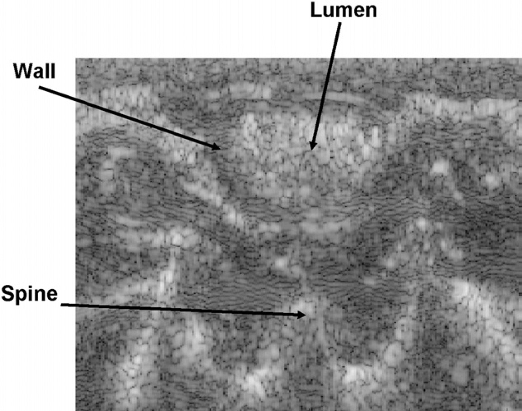
Fig. 2.
Typical ultrasound image view. Hypoechogenic circle around hyperechogenic bowel contents in the lumen represents the colon wall. The strain developed inside of the intestinal wall is normalized to the average strain estimated by the displacement of the spine relative to the surface of the transducer.

