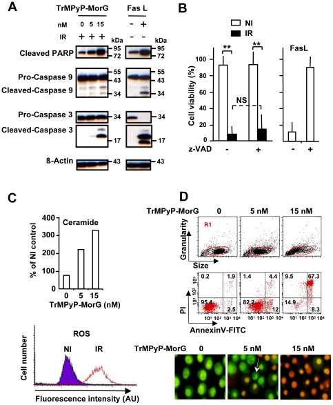
Figure 4. Cell death signaling triggered by PCT using TrMPyP-MorG conjugate.
Jurkat T cells (106/mL) were incubated with various TrMPyP-MorG concentrations for 15 min at 37°C and in the dark. After washing, cells were irradiated (IR) or not (NI), for 7.5 min. (A) Western blot analysis of cells 3 h post-irradiation. FasL (50 ng/mL) was used as a control for inducing apoptosis. (B) Jurkat T cells were incubated in the presence (+) or absence (−) of 20 µM of the pan-caspase inhibitor (z-VAD), then treated by TrMPyP-MorG (15 nM) and irradiated (IR) or not (NI). FasL-induced cell death was used as a control. Cell death was analyzed using MTT assay, 24 h after PCT. Results are means ± SD of 4 separate experiments (similar results were obtained with 5 nM TrMPyP-MorG), ** P<0.001, Anova test; NS, not significant. (C) Intracellular ceramide concentration was determined 3 h after PCT. Results are expressed as percentage of the ceramide levels in drug-treated controls without irradiation (mean of two separate experiments; similar results were observed 24 h post-PCT). Cellular ROS levels were analyzed using flow cytometry. Representative results after TrMPyP-MorG (5 nM) treatment and 15 min post-irradiation. (D) Three hours after irradiation, cells were stained with AnnexinV-FITC/PI (upper panel) or Syto13/PI (lower panel) and cell death was evaluated by flow cytometry (in gate R1, defined to exclude cell debris from the analysis) or by fluorescence microscopy, respectively. The arrow indicates a cell with classical features of apoptosis (reduction of cellular volume, nuclear fragmentation, PI exclusion).

