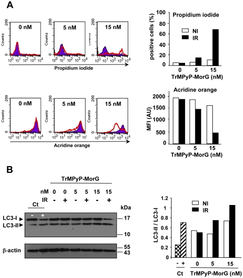
Figure 6. Induction of necrosis by TrMPyP-MorG conjugate.
Parental Jurkat T cells (106/mL) were incubated with TrMPyP-MorG (5 or 15 nM) for 15 min at 37°C in the dark. After washing, cells were irradiated (IR) or not (NI) by white light (7.5 min, 1.7 J/cm2) and incubated 3–4 h at 37°C in the dark. (A) The cell viability was evaluated using PI staining, whereas the formation of acidic vesicular organelles (AVO) was assessed by using acridine orange. Cells were analyzed by flow cytometry excluding cell debris. Left, representative histograms: NI cells, full histograms; IR cells, empty histograms. Right, means of two independent experiments. (B) Left, representative Western blot to detect the conversion of LC3-I to LC3-II. Right, densitometry analysis; results are means of two independent experiments. Control cells (Ct) were cultured for 2 days either in RPMI medium containing 10% FCS (−) or HBSS medium (without amino acids and FCS) (+), a condition known to induce autophagy and AVO formation.

