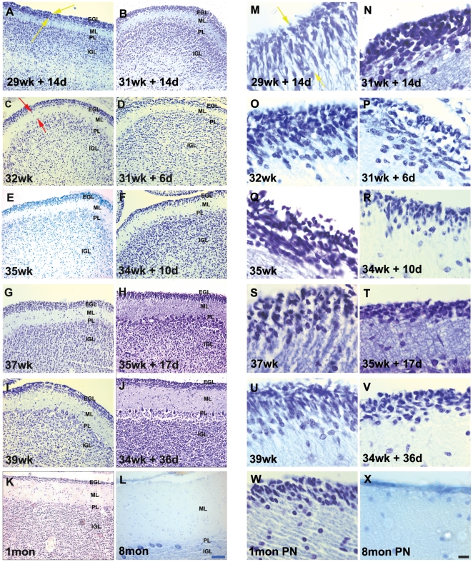
Figure 1. Cresyl violet stained sections of the developing human cerebellum.
(A–X) A greater EGL cell density and reduced EGL thickness were reported in preterms with ex-utero exposure, as compared to their age matched stillborn controls. ML thickness was increased in preterms with ex-utero exposure born after 34 weeks gestation, as compared to their age matched controls. Compare E with F, G with H and I with J. Abbreviations used - wk = number of gestational weeks, d = number of postnatal days, mon PN = postnatal months - born at term. EGL = external granular layer, ML = molecular layer, PL = purkinje cell layer, IGL = internal granular layer. Scale bar = 50 µm (A–L) and 20 µm (M–X).

