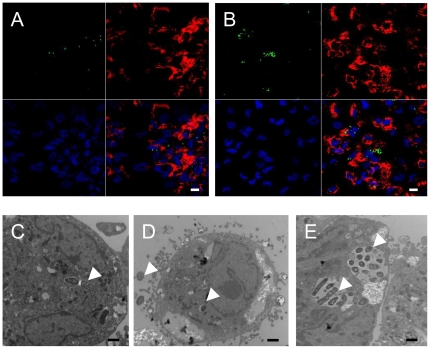
Figure 6. Survival and cytotoxic effects of EHEC strains within THP-1 macrophages.
Confocal microscopy (A and B) and Transmission Electron Microscopy (TEM) (C, D, and E) analysis of THP-1 macrophages infected with EHEC strains. THP-1 macrophages were infected with the 86-24 WT strain (A, C and D) and with the 86-24 Δstx2 isogenic mutant (B and E) at 1 h post-infection (A and C) and at 24 h post-infection (B, D and E). For confocal micrographs, THP-1 macrophages were infected with GFP-positive 86-24 WT and 86-24 Δstx2 isogenic mutant (green color). The actin cytoskeleton of cells was stained with TRITC phalloidin (red), and DNA was stained with DAPI (blue). Arrowheads indicate bacteria within THP-1 macrophages. Scale bar = 10 µm (A and B). Scale bar = 1 µm (C, D and E).

